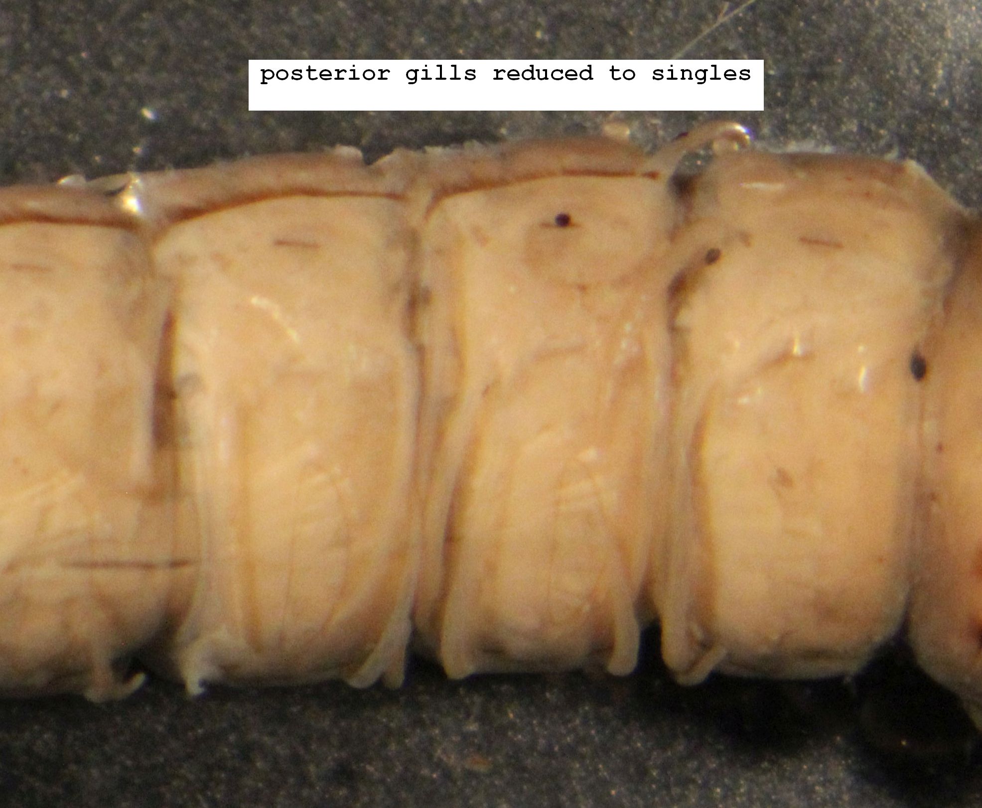
1. Most abdominal gills single (Fig. 1.1). . . . . . . . . . . . . . . . . . . . . . . . . . . . . . . . . . . . . . . . . 2
Most dorsal and ventral gills multiple, lateral gills sometimes single (Fig. 1.2). . . . . . . . . . . . . 4
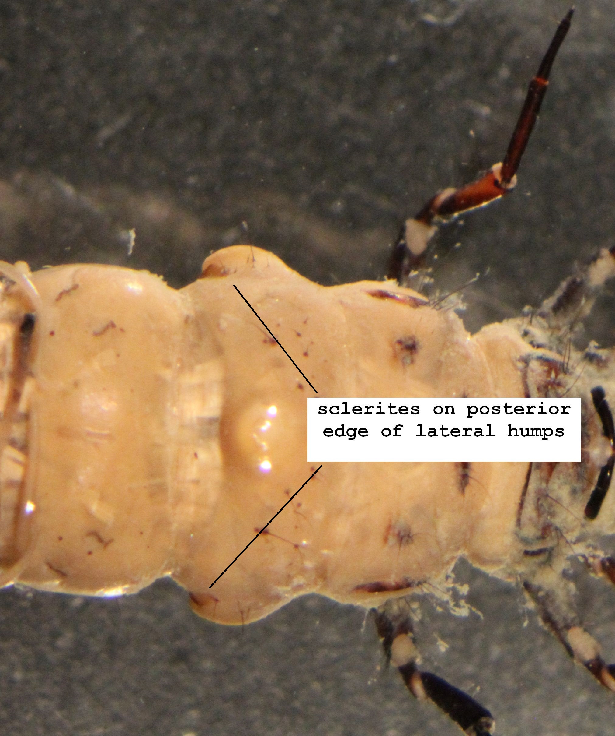
2. 1 or 2 long sclerite at posterior edge of base of each lateral hump of abdominal segment 1 (Fig. 1.3, Fig. 1.4)); sclerites often only lightly pigmented and difficult to see, but distinguishable by relatively shinier surfaces.. . . . . . . . . . . . . . . . . . . . . . . . . . . . . . . . . . . . 3
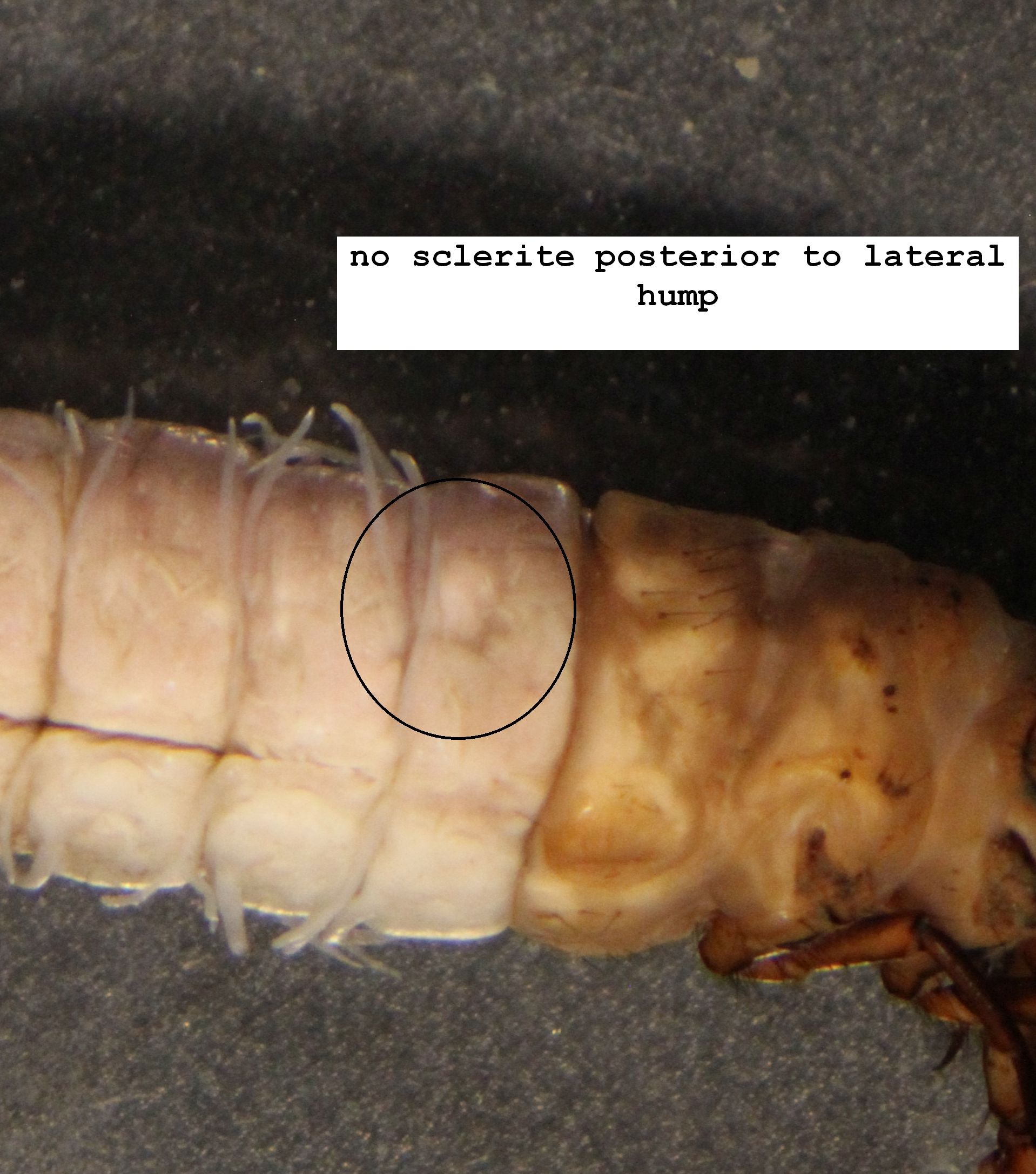
No sclerites adjacent to lateral humps on abdominal segment 1 (Fig 1.5). The two species of Pseudostenophylax, for Ohio, P. sparsus and P. uniformis are not clearly distinguished as larvae (Morse, et al., 2017). . . . . . . . . . . . . . . . . . . . . . . . . . . . . . Pseudostenophylax sp.
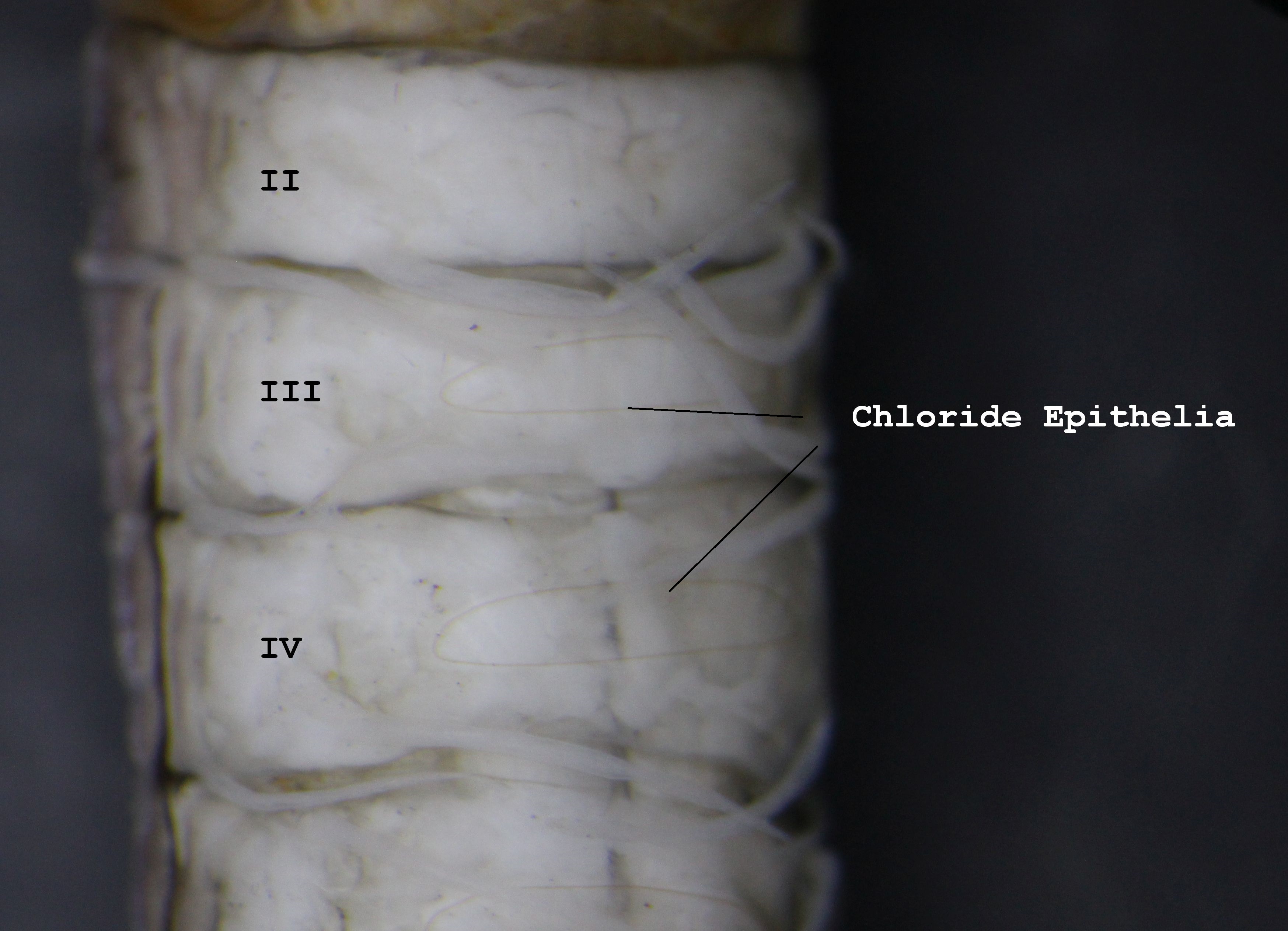
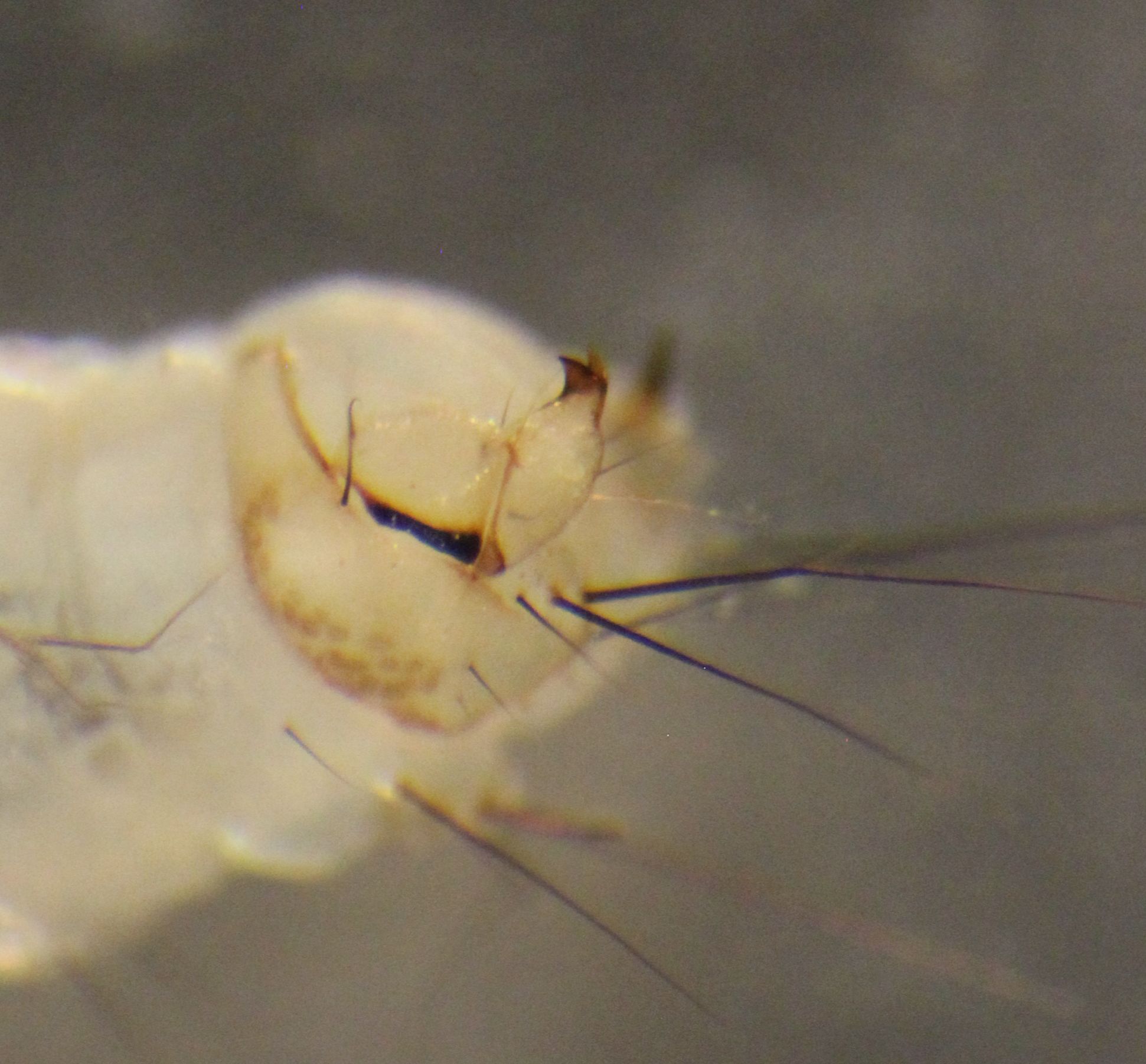
3. Metanotal sa1 sclerites fused (Fig. 1.6); abdominal sternum II with chloride epithelium (Fig. 1.7) (in which case abdominal segment IX with only single seta on each side of dorsal sclerite; case of wood or leaves in irregular outline. . . . . . . . . . . . . . . . Hydatophylax argus
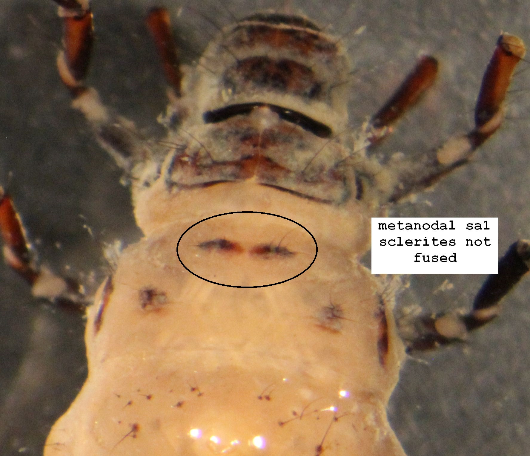
Metanotal sa1 schlerite not fused although often contiguous (Fig. 1.8), abdominal sternum II without cholride epithelium and abdominal segment IX with only single seta on each side of dorsal sclerite (difficult to see); case of twigs, gravel, or leaves, variously shaped, occasionally 3-sided. . . . . . . . . . . . . . . . . . . . . . . . . . . . . . . . . . . . . . . . . . . . . . . . . . . .Pycnopsyche
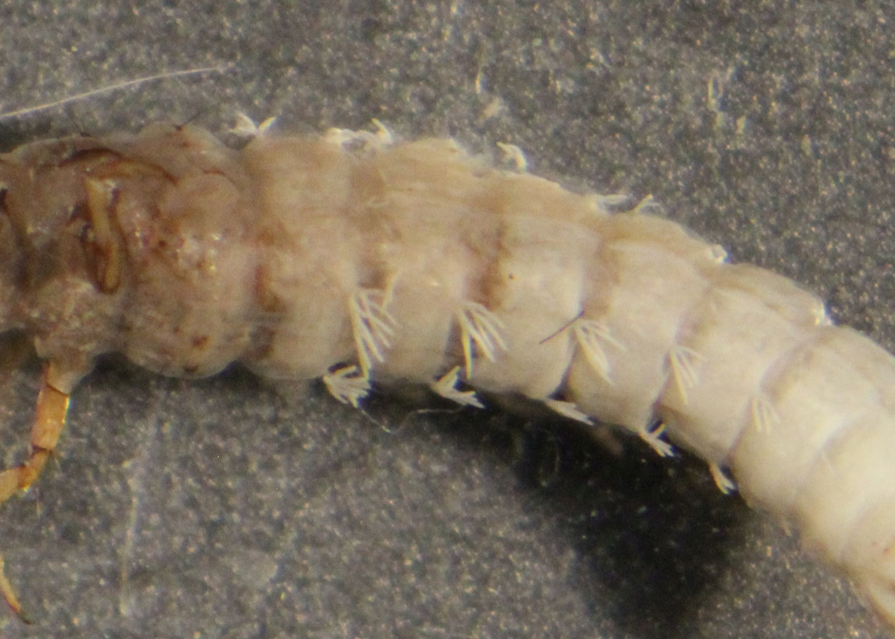
4. Most gills with 2 or 3 branches, none with more than 4 (Fig. 1.9). . . . . . . . . . . . . . . . . . . . 5
At least some gills with more than 4 branches (Fig. 1.10). . . . . . . . . . . . . . . . . . Ironoquia
5. Dorsum of head with 2 bands of contrasting color extending from coronal suture to bases of mandibles and/or narrowed posterior portion of frontoclypeal apotome with 3 light areas: 1 along each side and 1 at posterior extremity. . . . . . . . . . . . . . . . . . . . . . . . . . . . . . . . . . . 6
Dorsum of head lacking bands or other well-defined, contrasting areas, usually uniform in color or with prominent light or dark spots only at points of muscle attachment (Fig. 1.13). . . . . . 10
6. Dorsum of head with prominent dark bands on light background, lateral bands extended from coronal suture to base of each mandible, median band on frontoclypeus. . . . . . . . . . . . . . . 7
Dorsum of head lacking dark bands, but narrowed posterior part of frontoclypeus with 3 light areas; 1 along each side and 1 at posterior extremity. . . . . . . . . . . . . . . . . . . . . . . . . . . . . 9
7. Dark dorsal bands on head fussed at junction of coronal and frontoclypeal sutures to form U-shaped marking, pronotum with narrow dark band along anterior border and across dorsum; case of leaf pieces arranged transversely or longitudinally. . . Nemotaulius hostilis
Dark dorsal bands on head extended posterad beyond junction of coronal and frontoclypeal sutures to form V-shaped marking, pronotal markings variable. . . . . . . . . . . . . . . . . . . . . . . 8
8. Chloride epithelia absent dorsally (as well as dorsolaterally and ventraly) on several abdominal segments; rough tubular case of wood or leaf fragments, sometimes 3-sided, changed to fine gravel before pupation. . . . . . . . . . . . . . . . . . . . . . . . . . . . . . Halesochila taylori (Banks)
Chloride epithelia absent dorsally (present ventrally and sometimes dorsolaterally). . . . . . . . 9
9. Chloride epithelia present dorsally, laterally, and verntrally on most abdominal segments; case of plant and rock materials. . . . . . . . . . . . . . . . . . . . . . . . Asynarchus montanus (Banks)
Chloride epithelia absent dorsally, may be present dorsolaterally but always present ventrally on most abdominal segments. . . . . . . . . . . . . . . . . . . . . . . . . . . . . . . . . Limnephilus (in part)
10. Pronotum, especially anterior margin, and lateral sclerite of each anal proleg both with short, stout setae. . . . . . . . . . . . . . . . . . . . . . . . . . . . . . . . . . . . . . . . . . . . . . . . . . . . Frenesia
Pronotum usually (Fig. 1.14)(lateral sclerite of anal proleg always) without short, stout setae (Fig. 1.15). . . . . . . . . . . . . . . . . . . . . . . . . . . . . . . . . . . . . . . . . . . . . . . . . . . . . . . . . . . 11
11. Chloride epithelia present dorsally on at least some abdominal segments; case cylindrical, made of plant materials, sometimes 3-sided. . . . . . . . . . . . . . . . . . . . . . . . . . . . . . . . Anabolia sp.
Chloride epithelia absent dorsally on abdominal segments. . . . . . . . . . . . . . . . . . . . . . . . . 12
12. Prosternal horn extinding beyond head capsule to mentum of labium; case cylindrical, made of transverse, narrow, projecting pieces of plant material. . . . . . .. . . . Platycentropus radiatus
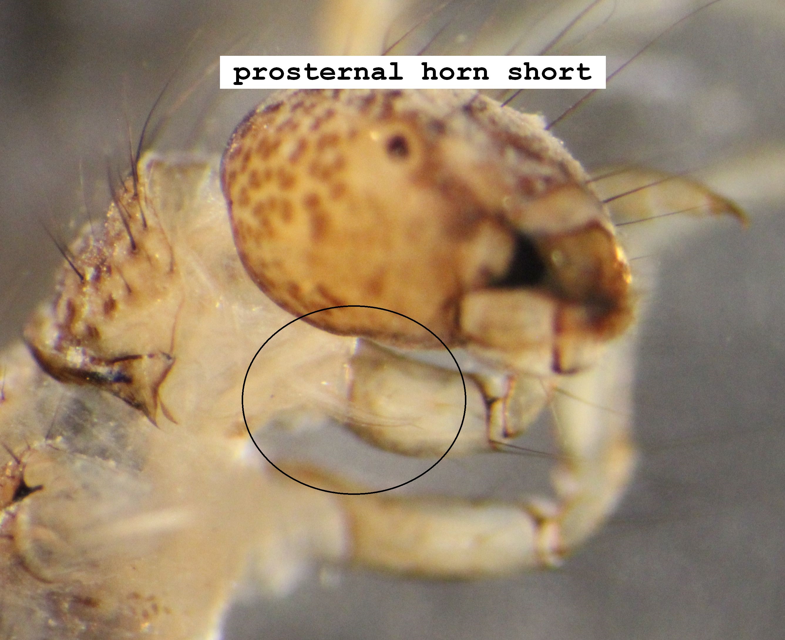
Prosternal horn extending only to distal edge of head capsule (Fig. 1.16); case with wide range of shapes and materials. . . . . . . . . . . . . . . . . . . . . . . . . . . . . . . . . . . Limnephilus (in part)
Key to Genera of Limnephilidae Nymphs
.JPG)
.JPG)